TOMSK,
May 2 – RIA Tomsk. Detectors
developed at Tomsk State University (TSU) will be installed on the
first X-ray microscope in Germany; it allow to look at the level of
atoms, and Tomsk detectors will help to reproduce the "picture".
In 2017, for this project they were put on for 300 thousand euros.
How high-tech science become profitable – in the material of RIA
Tomsk.
How
to make out molecules and atoms
Modern
microscopes allow to study objects with a dimension equal to the
wavelength of visible light radiation. It is about one micrometer
(one thousandth of a millimeter). To see smaller particles, it is
necessary to "highlight" them in a special way.
As
the head of TSU Laboratory for Functional Electronics, professor Oleg
Tolbanov explains, the role of such "highlighting" is
played by synchrotron radiation.
"The
synchrotron radiation wavelength – is one tenth of an angstrom
(0.00000001 millimeters), that is, objects of approximately this size
can be investigated. And what is 0.1 angstroms? These are the sizes
of the smallest molecules or very large atoms. This ultra-modern
microscope is being created now in Germany, in the
electron-synchrotron center DESY, one of the most powerful in the
world. In 2020, experiments should begin", – says Tolbanov.
This,
of course, will not be a desktop device, but a huge complex system
worth 1.5 billion euros, which heart – is a linear electron
accelerator that occupies a whole tunnel underground.
In
it, electrons are accelerated to very high energy – 17.5
gigaelectronvolts, then special undulators convert it into
synchrotron or x-ray coherent radiation. "Coherent means that
quanta have the same wavelength of radiation or the same energy", – explains Tolbanov.
But
to create radiation – is half the battle …
© предоставлено Олегом Толбановым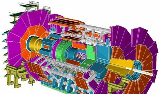
Linear particle accelerator
What
did Tomsk citizens made for this?
"To
get the "picture" obtained by X-ray illumination, detectors
needed – special receivers of synchrotron radiation, which this
information would reproduce in the form of pulses of electric current
in the counting mode of single quanta, that is, single "portions"
of energy, and then special equipment amplify them, digitize and
reproduce them on a computer monitor in the film version", – says the scientist.
The
equipment for converting electrical impulses exists, but fast-acting
quantum-sensitive detectors working in the right energy range were
not in the world – until they were made in Tomsk.
© предоставлено пресс-службой Томского госуниверситета
About 40 years ago radiophysicists of NIIPP and SPTI under the guidance of professor Stanislav Khludkov engaged in gallium arsenide properties modification and obtained new properties of semiconductor materials. The development of detectors began in 1993, the group was headed by Oleg Tolbanov.
"Synchrotron
radiation has a well-defined frequency, or quantum energy. We created
material which is sensitive to single external influences and allows
to register single quanta of X-ray radiation", – tells Tolbanov.
He
notes that the technology is rather complicated: "We introduce
one chromium atom per million gallium and arsenic atoms. Then put it
in the right place – and the properties of such material change
significantly. Thus, the ability to conduct electric current is
reduced by 10 billion times!".
The
functions of the detector can be roughly compared with a digital
camera.
"The
camera compresses the picture and projects it onto the receiving part
in the form of a compressed image. When playing this image needed to
be re-opened full screen. The device itself does not allow to do
this, but the circuit that works with this device allows it. You
insert the USB cable and see the image on the computer. This is
roughly the same with our detectors - only in other scales and in
energy range", – says the scientist.
And
the picture turns out color – unlike existing analogues on the
market, which reproduce only a black and white image.
© предоставлено Олегом Толбановым
The creation of one large detector takes three months, a small one - one. All production is in Tomsk. Part of the process takes place in a very clean room, where only one employee has access. When working, he puts on a special suit.
The
competitors of Tomsk development Tolbanov calls mainly Japanese firms
that have semiconductor material cadmium telluride (CdTe). But it is
extremely whimsical and expensive – more expensive than Tomsk in
dozens of times.
"In
terms of physical properties, the Japanese material also loses to us,
so the market prefers our material. This is the direction that we
really are ahead of the curve", – emphasizes the scientist.
For
one project in DESY TSU radiophysicists is planning to produce three
to five large area detectors and three to four of small ones a year,
in 2017 the volume of deliveries was 300 thousand euros. In general.
For a year TSU Laboratory for Functional Electronics earned almost 10
rubles for every ruble of budget financing.
What
quantum detectors can
Now
Tomsk detectors are used in leading scientific centers of the world
for conducting modern experiments in physics.
"In
the synchrotron centers (there are 59 of them in the world, two of
them in Russia, and the most powerful one in Grenoble), experiments
are being performed at very high pressures and very high
temperatures. Thus modeling the processes taking place in the bowels
of the Earth, in the bowels of stars. And our detectors are used for
performing such experiments", – says Tolbanov.
The
development of Tomsk radiophysicists allows to solve the problems of
digitalization of images in X-ray and gamma-rays. For example, their
colleagues from Joint Institute for Nuclear Research (Dubna) bought
an American-New Zealand microtomograph, pulled out silicon detectors
and replaced them with Tomsk ones.
And
at University of Freiburg (Germany) in collaboration with Tomsk
residents was made a new X-ray machine. Tolbanov shows a picture of a
mouse head, received with the help of it, where the hard and soft
tissues, blood vessels are clearly drawn, and even some point,
perhaps a tumor, is visible.
"Doctors
look at this image and say: "Is this a tomography?". When
they get to know that this is just a projection shot, they are
surprised. Experiments are only going on in mice, but it's just a
matter of time when such technologies reach people", – notes the
scientist.
© предоставлено Олегом Толбановым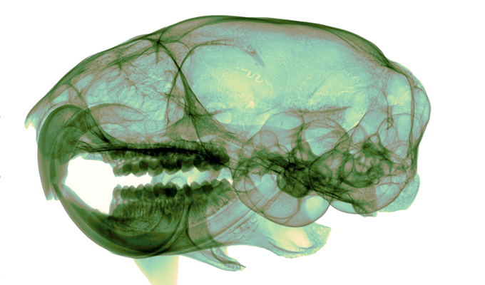
X-ray picture of the mouse's head